MEDICAL IMAGING
Electrical Impedance Tomography
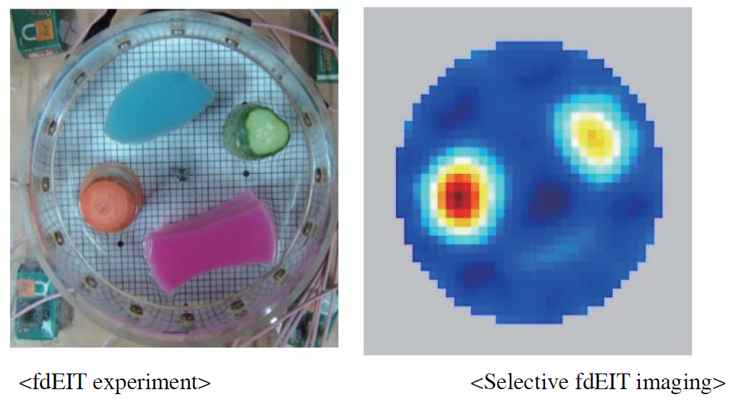
Considering the fundamental drawback of the static EIT in recovering the absolute admittivity image at a certain frequency, we focus on a difference imaging method using a currently available EIT system. We are particularly interested in the frequency-difference EIT (fdEIT) since it may provide spectroscopic admittivity images without requiring a time reference data. Noting that non-negligible susceptivity values of biological tissues are attributed to thin cell membranes, we analyze the role of the membrane in terms of the sensitivity of the complex voltage data in fdEIT.
We perform numerical simulations and phantom experiments of a two-dimensional imaging object containing an anomaly with a thin insulating membrane. The results provide better understanding on the role of the thin membrane to the sensitivity of a multi-frequency current–voltage data.
|
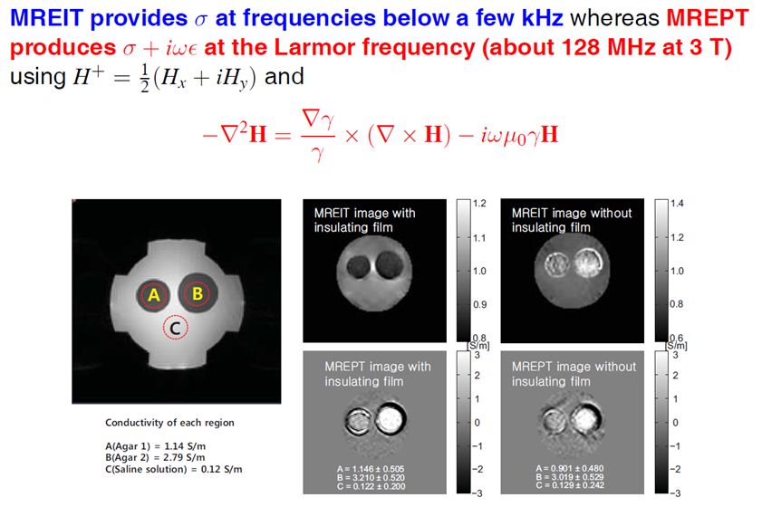
Magnetic Resonance EIT
Professor Jin Keun Seo developed the first irrotational MREIT (Magnetic Resonance Electrical Impedance Tomography) providing the most advanced electrical tissue property imaging. His research team which included E J Woo and O Kwon had carried out mathematics-oriented multidisciplinary research that combined knowledge and techniques starting from mathematical modeling and analysis to algorithm development and human experiments, and obtained an advanced source technology providing
the highest spatial resolution of admittivity distribution to date. LV contours in Ultrasound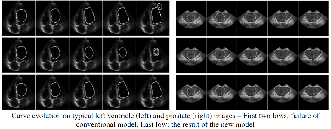
Segmentation of a target object in the form of a closed curve has many potential applications in medical imaging because it provides quantitative information related to its size and shape including measurement of the prostate in urology, detection and measurement of liver tumors, volume change of the left ventricle for examples. However, ultrasound image segmentation for boundary delineation of the target object is a very difficult task due to its inherent drawbacks including uncertainty of segmentation boundary caused by speckle noise, relatively low SNR and low contrast.
The new model may be combined with various fitting terms to enhance segmentation results. Numerical experiments show that the proposed model more effectively detects the target object in the form of a closed contour from low SNR ultrasound images, comparing the conventional arc length minimizer. Dental CT : Metal Artifacts Reduction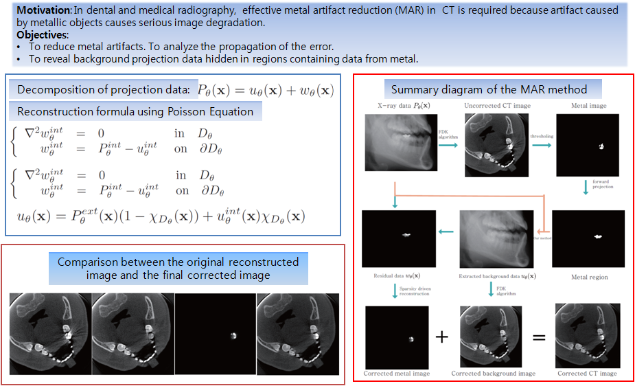
There is increasing demand in the field of dental and medical radiography for effective metal artifact reduction (MAR) in computed tomography (CT) because artifact caused by metallic objects causes serious image degradation that obscures information regarding the teeth and/or other biological structures.
This research presents a new MAR method that uses the Laplacian operator to reveal background projection data hidden in regions containing data from metal. In the proposed method, we attempted to decompose the projection data into two parts: data from metal only (metal data), and background data in the absence of metal. Removing metal data from the projections enables us to perform sparsity-driven reconstruction of the metal component and subsequent removal of the metal artifact. The results of clinical experiments demonstrated that the proposed MAR algorithm improves image quality and increases the standard of 3D reconstruction images of the teeth and mandible. Quantitative susceptibility mapping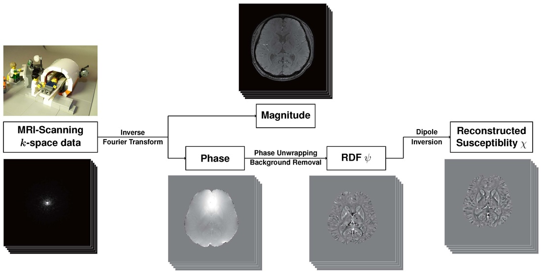
Quantitative susceptibility mapping (QSM) is a new medical imaging technique that can visualize magnetic susceptibility, changes of which in tissue indicate various disease processes involving iron transport. The inverse problem of QSM is to recover the susceptibility distribution of the human body from the measured local field that is expressed by the convolution of the susceptibility distribution with the magnetic field generated by a unit dipole. The inverse problem is ill-posed due to the presence of zeros at a cone in the Fourier representation of the unit dipole kernel. Reconstruction methods have been greatly improved to give better recovery of tissue susceptibility data for QSM, and various clinical applications have been pursued.
|